New scientific techniques are revealing the intricate role that proteins play in photosynthesis.
Despite being discovered almost 300 years ago, photosynthesis still holds many unanswered questions for science, particularly the way proteins organise themselves to convert sunlight into chemical energy and, at the same time, protect plants from too much sunlight.
Now a collaboration between researchers at Leeds and Kobe University in Japan is developing a novel approach to the investigation of photosynthesis.
“We now have a testbed and a suite of advanced imaging tools that will reveal the molecular workings of photosynthesis.”
Using hybrid membranes that mimic natural plant membranes and advanced microscopes, they are subjecting photosynthesis to nanoscale investigation – the study of life at less than one billionth of a metre – to reveal the behaviour of individual protein molecules.
Dr Peter Adams, Associate Professor in the School of Physics and Astronomy at Leeds, who supervised the research, said: “For many decades scientists have been developing an understanding of photosynthesis in terms of the biology of whole plants. This research is tackling it at the molecular level and the way proteins interact.
“A greater understanding of photosynthesis will benefit humankind. It will help scientists identify new ways to protect and boost crop yields, as well as inspire technologists to develop new solar-powered materials and components.”
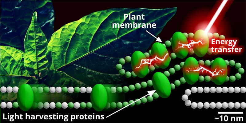
Photosynthesis happens when photons or packets of light energy cause pigments inside light-harvesting proteins to become excited. The way that these proteins arrange themselves determines how the energy is transferred to other molecules.
It is a complex system that plays out across many different pigments, proteins, and layers of light-harvesting membranes within the plant. Together, it regulates energy absorption, transfer, and the conversion of this energy into other useful forms.

Light is absorbed by light-harvesting proteins in the plant membrane. The energy excites the proteins and the way they arrange themselves determines the way energy is transferred through the plant. Credit: Sophie Meredith.
To understand this intricate process, scientists have been using a technique called atomic force microscopy, which is capable of revealing components of a membrane that are a few nanometres in size.
The difficulty is that natural plant membranes are very fragile and can be damaged by atomic force microscopy.
The team at the University of Leeds used the hybrid membrane and subjected it to atomic force microscopy and another advanced visualisation technique called fluorescence lifetime imaging microscopy, or FLIM.
PhD researcher Sophie Meredith, also from the Leeds' School of Physics, is lead author of the paper. She said: “The combination of FLIM and atomic force microscopy allowed us to observe the elements of photosynthesis. It gave us an insight into the dynamic behaviors and interactions that take place.
“What is important is that we can control some of the parameters in the hybrid membrane, so we can isolate and control factors, and that helps with experimental investigation.
“In essence, we now have a testbed and a suite of advanced imaging tools that will reveal the molecular working of photosynthesis.”
Further information
The research was supported by the Royal Society, the Biotechnology and Biological Sciences Research Council, the Engineering and Physical Sciences Research Council, the Medical Research Council and the Japan Society for the Promotion of Science.
For media enquiries, contact David Lewis in the press office at the University of Leeds via d.lewis@leeds.ac.uk.