A groundbreaking operation to remove a patient’s brain tumour through her eye socket has been performed in Leeds.
Ruvimbo Kaviya, 40, is the first person in the UK to undergo the procedure, which involves a flexible tube and camera inserted through the eye socket.
Surgeons affiliated to the University carried out the procedure at Leeds General Infirmary (LGI). Mr Asim Sheikh is part of the Leeds Institute of Medical Research (LIMR) and is a Consultant Skullbase and Neurovascular Neurosurgeon. Mr Jiten Parmar from the School of Dentistry and the School of Medicine at Leeds is a Consultant in Maxillofacial Surgery and completed his medical degree at the University of Leeds.
Ruvimbo, who received her diagnosis in 2023, had been experiencing persistent headaches for months, initially managed with pain relief. But the pain had intensified, with spasm-like sensations that led her to believe she had a toothache. A visit to her dentist revealed no issues, prompting her to seek help at the emergency department at LGI.
After an MRI, she was diagnosed with meningiomas on the right side of the back of her brain and left side of her eye - pressing on the nerve surrounding her eye. It was in the area called cavernous sinus which is difficult to access and is considered somewhat inoperable.
Ruvimbo said: “The diagnosis came as a shock. It was an incredibly stressful and overwhelming time. The pain was severe, and managing my medication was difficult. When I was told about the surgery, I stayed optimistic as the tumour was growing. Going ahead with the procedure was the best option for me.”
Minimally invasive
The procedure represents a shift in how skull base tumours are treated. Unlike traditional surgery, which often requires large incisions, the removal of parts of the skull and extended recovery time, this innovative technique is minimally invasive. Surgeons accessed the tumour through a small, 1.5cm incision on the side of Ruvimbo’s eyelid.
“I was amazed by the recovery,” Ruvimbo said. “I was only in the hospital for two days, with no side effects or swelling. I feel perfectly fine now. I am deeply grateful to Mr Sheikh, Mr Parmar, and the entire team—they reassured me throughout the process.”
Surgery on tumours at the base of the skull has traditionally required an open craniotomy, a procedure involving significant trauma to the surrounding tissues and a lengthy recovery. The endoscopic trans-orbital approach offers a safer alternative with a faster healing process.
Mr Sheikh said: “This technique allows us to remove tumours without opening the skull or having to retract or compress the brain. The minimally invasive nature of the procedure significantly reduces trauma, enabling patients to recover faster with minimal visible scarring.”
Mr Parmar said: “The ability to work collaboratively with maxillofacial and neurosurgical teams, using cutting-edge 3D planning, has been a game changer. This partnership allows us to precisely target the tumour, ensuring safer outcomes for patients.”
The success of this surgery was underpinned by innovative 3D planning led by Lisa Ferrie, Biomedical Engineer and 3D Planning Service Lead at Leeds Teaching Hospitals NHS Trust. The 3D model was used to perform steps of surgery on the life size model, prior to actual surgery taking place.
“When the surgical team approached me, we used scans of Ruvimbo’s brain and skull to create a 3D replica model,” Lisa explained. “This technology enabled the team to study her anatomy in detail and prepare for the procedure with unparalleled accuracy. Seeing the model and knowing it contributed to this groundbreaking surgery is incredibly rewarding.”
Further information
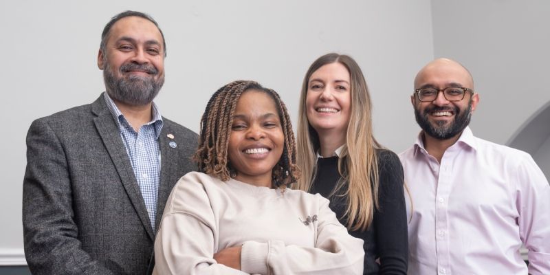
Picture: Mr Asim Sheikh, Ruvimbo Kaviya, Lisa Ferrie and Mr Jiten Parmar. Credit: PA Media