A tiny magnetic robot which can take 3D scans from deep within the body and could revolutionise early cancer detection, has been developed by researchers.
The team, led by engineers from the University of Leeds, say this is the first time it has been possible to generate high-resolution three-dimensional ultrasound images taken from a probe deep inside the gastrointestinal tract, or gut.
It paves the way to a transformation of the diagnosis and treatment of several forms of cancer by enabling ‘virtual biopsies’ — non-invasive scans that provide immediate diagnostic data, allowing doctors to detect, stage, and potentially treat lesions in a single procedure: eliminating the need for physical biopsies.
The perhaps surprising key to the team’s success was the use of a little-known 3D shape, the oloid, which gave the magnetic medical robot a previously impossible range of motion – the roll – essential for precise navigation and imaging inside the body.
A paper, published yesterday (March 26) in Science Robotics, explains how the team integrated the oloid shape, and its unique rolling motion, into a new magnetic flexible endoscope (MFE). They equipped it with a small, high-frequency imaging device to capture detailed 3D images of internal tissues.
This technology was developed through a collaboration between engineers, scientists, and clinicians from the University of Leeds, the University of Glasgow, and the University of Edinburgh. Leeds led the robotics development and integration of the probe, while Glasgow and Edinburgh provided the ultrasound probe and led the imaging component.
Pietro Valdastri, Professor and Chair in Robotics and Autonomous Systems and Director of the STORM Lab, coordinated the research behind this paper. He said: “For the first time, this research enables us to reconstruct a 3D ultrasound image taken from a probe deep inside the gut - something that has never been done before.
“This approach enables in-situ tissue analysis and diagnosis of colorectal cancer, with immediate results. The process of diagnosing colorectal cancer currently requires a tissue sample to be removed, then sent to a lab, with results taking from one to three weeks.”
The imaging device – a 28 MHz micro-ultrasound array – creates a high-resolution, 3D reconstruction of the area it scans. From this virtual reconstruction, clinicians can make cross-sectional images that mimic those generated by a standard biopsy, in which a sample of tissue is sliced into thin layers and placed on a slide to be examined under a microscope.
High frequency, or high-resolution ultrasound, is different to the ultrasound most of us are familiar with, used to look at a foetus or internal organs. The high frequency/resolution ultrasound probe used in this study enables users to see features on a microscopic level, down to tissue layer level detail.
While 3D ultrasound can already be performed in blood vessels and in the rectum, this work opens the possibility for 3D scans to be performed deeper in the gastrointestinal tract.
For the first time, this research enables us to reconstruct a 3D ultrasound image taken from a probe deep inside the gut - something that has never been done before.
Postgraduate researcher Nikita Greenidge, a member of Leeds’ STORM Lab, in the School of Electronic and Electrical Engineering, is lead author of the paper. She said: “By combining our advanced robotics with medical ultrasound imaging, we take this innovation one step ahead of traditional colonoscopy, allowing doctors to diagnose and treat in a single procedure — eliminating the wait between diagnosis and intervention. This not only makes the process more comfortable for patients but also reduces waiting times, minimises repeat procedures, and alleviates the anxiety of waiting for potential cancer results.”
She added: “Colorectal cancer is one of the leading causes of cancer-related deaths in the UK and globally, but if detected early it is highly treatable. This research presents a new approach that could significantly improve early diagnosis with a minimally invasive approach and could also, in future, facilitate targeted ultrasound-triggered drug delivery for more effective treatment.”
The research was funded by UKRI Engineering and Physical Sciences Research Council (EPSRC), the European Commission (EC), the European Research Council (ERC), and the NIHR Leeds Biomedical Research Centre. It found that using the oloid – a shape formed by two intersecting perpendicular circles – significantly enhanced the dexterity, diagnostic capabilities and autonomy of magnetic flexible endoscopes, and magnetic medical robots overall.

Caption: The 3D-printed oloid. Credit: STORM Lab, University of Leeds
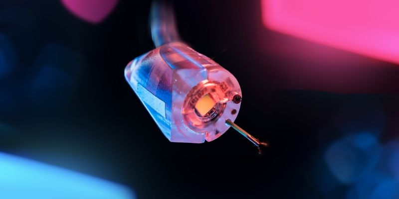
The oloid magnetic endoscope (OME) was 3D printed out of resin, measuring just 21 mm in diameter – around the size of a 1p coin - meaning the robot could still roll but was a practical size and design for clinical applications like colonoscopy. Its movement was tested on a range of surfaces simulating the structures of the colon, oesophagus and stomach.
To advance the technology toward human trials, the team first conducted tests in an artificial colon, followed by studies in pigs, a necessary step in meeting regulatory requirements for medical device approval. They used a robotically controlled external permanent magnet, a platform previously developed at Leeds which enables both joystick and autonomous control of the OME. Navigation was assisted by images from an embedded camera and a magnetic localisation system. The results demonstrated that the system could:
- Successfully perform controlled rolling and sweeping motions inside the colon.
- Generate high-resolution 3D ultrasound scans for accurate diagnosis.
- Identify lesions in gastrointestinal tissue, showcasing its potential for advanced medical imaging and early disease detection.
Ms Greenidge said while this research had been conducted in the colon, the rolling properties of the oloid shape could be applied to a variety of magnetic medical robots, potentially expanding its applications to other areas of the body.
This research presents a new approach that could significantly improve early diagnosis with a minimally invasive approach.
The team will now set about collecting all the data that will allow them to conduct human trials, which they hope could start in 2026, as the Leeds platform for robotic colonoscopy without ultrasound capabilities is already undergoing human trials and being commercialised by Atlas Endoscopy, a Leeds-based company formed by the STORM Lab.
The science of magnetic robots
Magnetic fields are ideal for medical applications as they pass harmlessly through human tissue, enabling the remote manipulation of tiny surgical robots. Controlled rolling and sweeping motions are essential for precise navigation and imaging inside the body. However, it is impossible to make cylindrical robots roll, using an external magnetic field.
Cylindrical magnetic robots can only achieve five degrees of freedom – the ways an object can move. This was considered a limitation: 3D scans weren’t possible without the rolling motion. Although gravity would cause a cylinder or sphere to roll down a slope, it’s not possible to make them roll using external magnetic forces. Utilising the oloid has solved that problem as its unique geometry naturally facilitates a meandering rolling motion that couples the roll to the up-down and side-to-side rotations. Because the oloid does not have symmetry around a central axis, the external magnet can apply a torque – or twisting force – inside the body, in two directions, to bring about the rolling motion.
Ms Greenidge said: “Our findings suggest new possibilities for interdisciplinary approaches to medical robotics, showing how mathematical principles like simple geometry can solve real-world healthcare challenges.”

Caption: External permanent magnet mounted onto a robotic arm to control the magnetic endoscope. Credit: STORM Lab, University of Leeds
Professor Sandy Cochran, Centre for Medical and Industrial Ultrasonics at the University of Glasgow, who led the ultrasound component of the study, said: “Ultrasound imaging is safe, inexpensive and can be deployed exactly where it’s needed. Through this collaborative approach, linking medical ultrasound imaging and cutting-edge robotics, we hope to help bring about transformative changes in cancer diagnosis, treatment, and patient management.”
The team believe the advancements they have made could lead to a fundamental change in endoscopy, where endoscopists can focus on critical diagnostic and therapeutic decisions while autonomous systems handle routine navigation and tasks.
They also believe the OME’s enhanced dexterity and diagnostic capabilities could help address gender disparities in colonoscopies, as standard flexible endoscope procedures tend to be more challenging in women, leading to higher rates of incomplete procedures.
Jane Nicholson, Executive Director of Research at EPSRC, said: “Progress from cutting-edge technology developments is enabling the development of rapid, non-invasive solutions that have the potential to revolutionise cancer diagnosis and treatment.
“By improving the precision and control of procedures for high-incidence cancers such as colorectal cancer, the efforts of this interdisciplinary team could lead to significant advancements in cancer detection and treatment.”
Further Information
The paper ‘Harnessing the oloid shape in magnetically driven robots to enable high-resolution ultrasound imaging’ was published in Science Robotics March 26, 2025.
For more information or to arrange interviews, email press officer Deb Newman via d.newman@leeds.ac.uk and pressoffice@leeds.ac.uk